
 There have been many advances in ultrasound technology in the past 20 years however, only three have had a significant affect on the quality of an ultrasound image.
They are: Tissue Harmonics Imaging, Compound Imaging, and Speckle Reduction Imaging*
Learning how to use these technologies is an important step in getting the best image quality out of your ultrasound machine.
*It is important to note that this blog will use the technologies common name as listed above. Every ultrasound manufacturer offers these technologies but call them by a different name. We will place as many known alternate names for each technology in parenthesis next to the common name.
Tissue harmonic imaging (THI, HARM, TI)
Is the most common advanced imaging technology that is available on new ultrasound systems and about 90% of used or refurbished ultrasound systems.
It creates images that are derived solely from the higher frequency, second harmonic sound produced when the ultrasound scanner pulse passes through tissue within the body. Tissue harmonics uses various techniques to eliminate the echoes arising from the main transmitted ultrasound scanner beam (“the fundamental frequencies”), from which conventional images are made. Once the fundamental frequencies are eliminated, only the harmonic frequencies are left for image formation.
Benefits of using Tissue harmonic imaging
The major benefit of tissue harmonic imaging is the reduction of image artifacts. Tissue harmonic images often exhibit:
- An improvement in contrast resolution (improved signal-to-noise)
- A less noisy images, making cysts appear clearer and improving visualization of pathologic conditions and normal structures
- Decreased scatter from the body wall
- Reduced artifacts from weak echoes such as side lobes, scatter, and grating lobes
- Less near field haze from body wall reverberations
When to use Tissue harmonic imaging
Tissue harmonics is very helpful when scanning any patient where there is a lot of ultrasound noise. For example, a patient with severe lower extremity swelling and soft tissue edema presents for a venous insufficiency evaluation. Ultrasound examination of the great saphenous vein below the knee is technically difficult due to the amount of enhanced through transmission (noise) caused by the amount of fluid present in the soft tissue. Turning on tissue harmonics helps to reduce this artifact, enabling imaging of accurate anatomy.
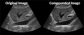
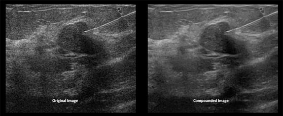
Compound Imaging (CrossXBeam, CRI, SonoCT. iBeam, OmniBeam, XView, SonoMB, ApliPure, Spatial Compounding)
Is a technology that acquires multiple frames from different steering angles and combines them into one image.
 Traditionally, transducers send and receive ultrasound signals in a single “line of sight.” This means it sends a sound signal perpendicular to the probe head, then listens for the returning echo.
 Compound imaging sends signals at multiple angles which and then displays a single image captured from multiple angles, eliminating artifacts.
Below is an ultrasound example of a "traditional" and of a compounded image.
Benefits of using compounding:
Compound imaging increases image resolution, eliminate artifacts, shadows, and increase edge detail.
How to use compounding:
Manufacturers offer compounding as an on/off setting or with selectable levels (the number or level typically refers to how many lines of sight the transducer is using… 3, 5, 7, etc.)
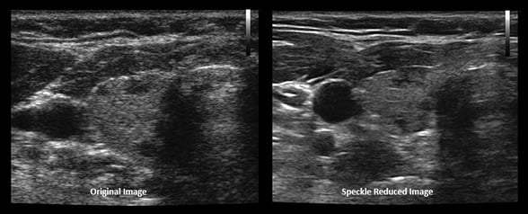
Speckle Reduction Imaging (SRI, uScan, XRES, TissuePure)
Speckle, the "dots" in an ultrasound image, represent an interference pattern caused by ultrasound waves scattering off small particles in the material or tissue being scanned.
Speckle Reduction Imaging, is an algorithm that provides a significant reduction in speckle, smoothing regions where no feature or edges appear while maintaining or enhance edges and borders. It increases contrast resolution by increasing the signal to noise ratio.
Benefits of using Speckle Reduction:
- The algorithm does not eliminate any information, so diagnostic criteria is preserved.
- The image quality improvements will help to improve consistency in diagnosis, reduce patient and operator dependence and may ultimately improve diagnostic accuracy and confidence and increase patient throughput.
How to use Speckle Reduction:
Most manufacturers offer selectable levels of Speckle Reduction. The lowest level reduces small amounts of artifact and lightly enhances tissue. The highest level can make the image look over-processed.
Depending upon the manufacturer, this is can be either a pre-processing or post-processing technology. When it is available as a post-processing technology you can adjust its level after the image is frozen.
Below is an ultrasound example of a "traditional" and of a "Speckle reduced" image.
With some practice, you’ll quickly become an expert on optimizing your ultrasound image.